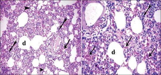
Figure 4.

Photomicrographs of the lung sections of experimental Group II showing cellular infiltrates (bifid arrow) and the alveoli were full of cellular infiltrates and alveoli became collapsed (arrowhead); others became irregular air spaces (d). Extravasated red blood cells (arrow) in the interalveolar spaces as well as the interstitial tissue (H and E, ×200, ×400)
