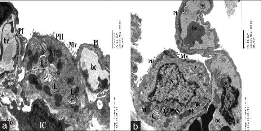
Figure 8.

Electron micrographs of ultrathin section of control group showing a part of patent alveolus lined with type I pneumocyte (PI) and type II pneumocytes(PII) with few short microvilli(Mv) on the surface, (a) euchromatic nucleus(N), characteristic lamellar bodies (L) and multiple mitochondria(M) (b). The interalveolar septum contains blood capillaries (bc) interstitial cell (IC) and some collagen fibers (Co) (a&b)
