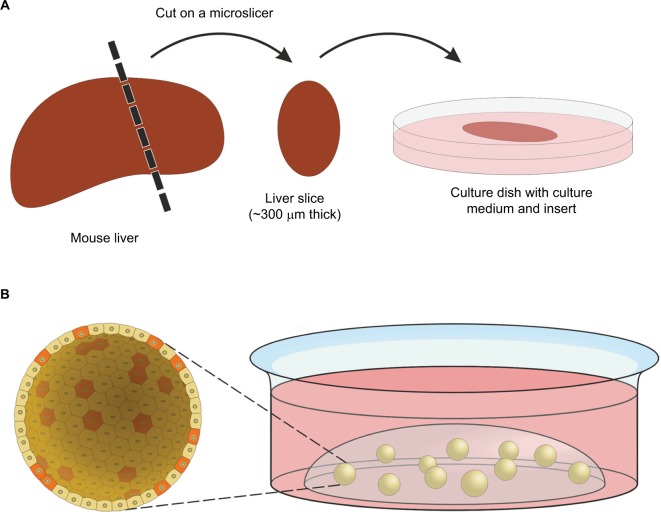
Figure 2.
Organotypic tissue slices and organoids.
Notes: (A) Preparation of an organotypic tissue slice. After cutting, slices can be cultured in cell-culture media for several days. Special inserts in culture dishes ensure that the slices are close to the surface of the culture medium for a sufficient oxygen supply. (B) Organoid culture, stem cells provided with stem cell, and niche factors form organoids in a three-dimensional matrix (in general hydrogel). A section through a spherical organoid (up to 500 µm in diameter) is enlarged so that single cells become visible. Stem-like cells are shown in red and differentiated cells in yellow. Cells in organoids form a flat structured surface outward. Internally, they build a lumen in which dead cells accumulate after some time.

