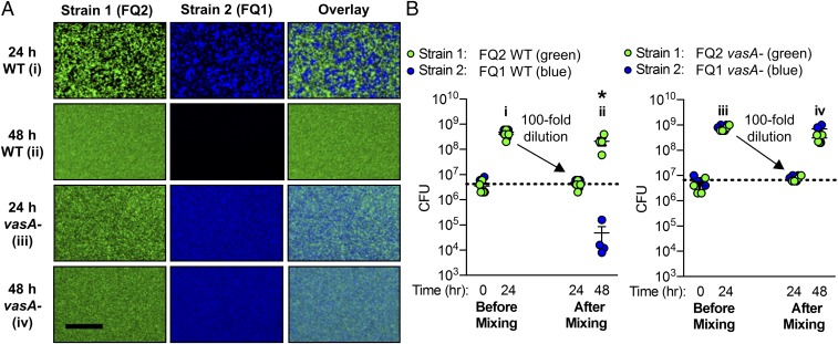
Fig. 6.
Spatial separation of lethal strains is not stable in the presence of physical mixing. (A) Fluorescence microscopy images for pairwise coincubations with FQ1 and FQ2 wild-type (WT) and vasA_2 mutant (vasA-) strains taken at 24 and 48 h (A); an overlay of images from columns 1 and 2 is shown in column 3; image will be light blue when strain 1 (green) and strain 2 (blue) cells are present in the same pixel. (Scale bar, 25 µm.) (B) Corresponding CFU counts for each coincubation spot. Coincubations were set up in the standard method (“before mixing”) and at 24 h were resuspended, mixed, diluted 100-fold, and spotted onto fresh LBS plates for the remainder of the assay (“after mixing”). Asterisk indicates P < 0.01 (Student’s t test) indicating a statistically significant decrease in a strain’s CFUs at 48 h compared with 24 h for each coincubation. All experiments were performed at least three times and a representative experiment is shown (n = 4).

