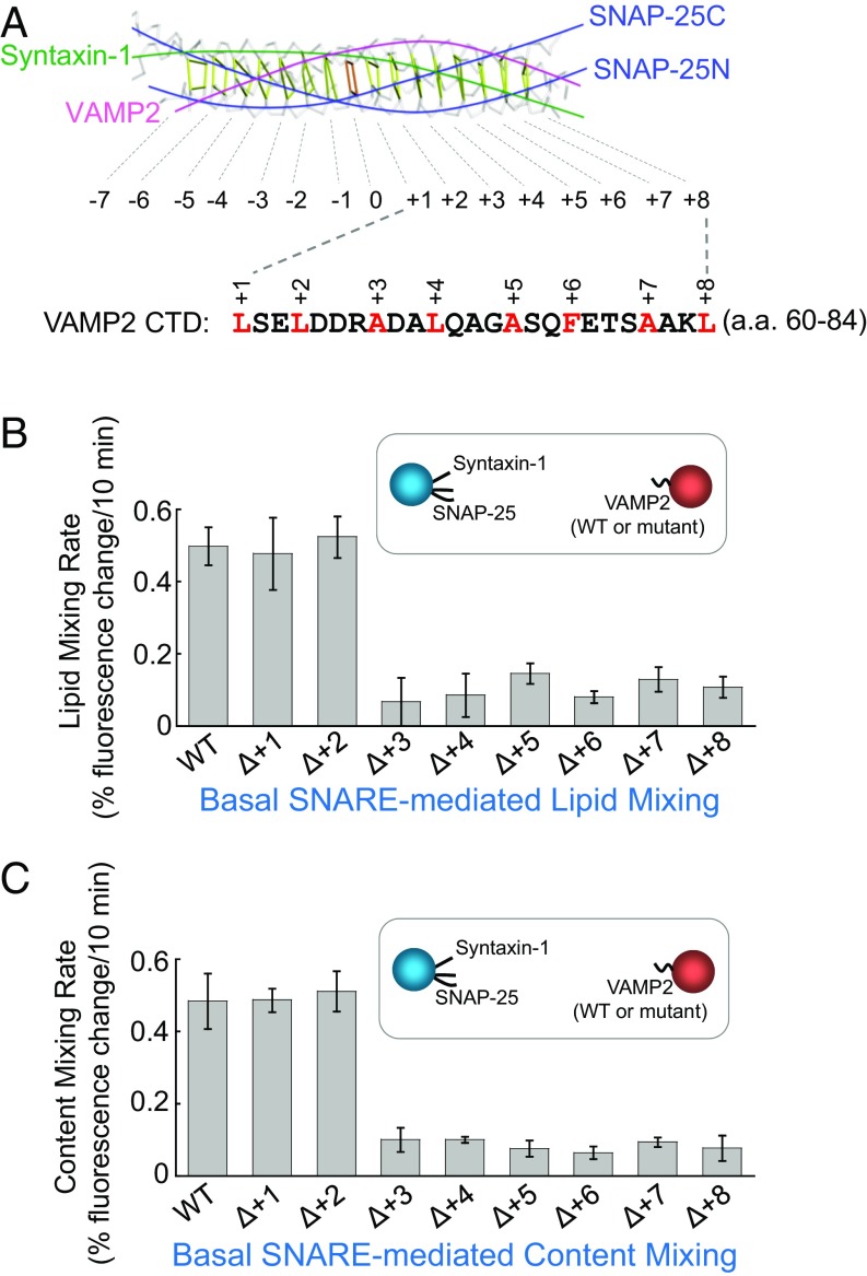
Fig. 1.
The +1 and +2 layers of the v-SNARE are dispensable for SNARE-mediated liposome fusion. (A, Top) Backbone view of the SNARE core bundle with individual layers indicated. (A, Bottom) The sequence of VAMP2 CTD (amino acids 60–84). Layer residues are numbered and highlighted. The model is based on the crystal structure of the synaptic SNARE complex (PDB ID code 1SFC) (8). (B) Initial lipid-mixing rates of the liposome fusion reactions reconstituted with WT synaptic exocytic t-SNAREs and WT or mutant VAMP2. In the layer deletion mutants, the layer residues were individually removed. Each fusion reaction contained 5 μM t-SNAREs, 1.5 μM v-SNARE, and 100 mg/mL of the macromolecular crowding agent Ficoll 70. Data are presented as the percentage of fluorescence change per 10 min. Error bars indicate SD. (C) Initial content-mixing rates of the above liposome fusion reactions.

