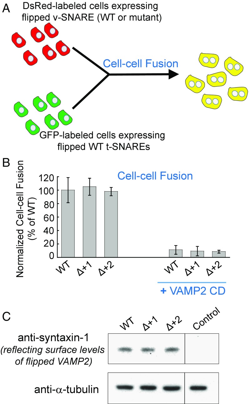
Fig. 2.
The +1 and +2 layers of the v-SNARE are not required in a flipped SNARE-mediated cell-cell fusion. (A) Diagram illustrating the cell-cell fusion assay. (B) COS-7 cells expressing flipped t-SNAREs were labeled with GFP while cells expressing flipped v-SNAREs were labeled with DsRed. After incubation, the fused cells (displaying both GFP and DsRed) were measured by flow cytometry. In the negative control, 20 μM VAMP2 cytosolic domain (CD, amino acids 1–95) was added at the beginning of the fusion reactions. Data are presented as normalized percentage of cell-cell fusion driven by WT-flipped SNAREs. Error bars indicate SD. (C) Immunoblots showing the surface expression levels of WT or mutant-flipped VAMP2. Since VAMP2 proteins could not be efficiently biotinylated (15), surface levels of VAMP2 were measured indirectly through its binding to t-SNAREs. COS-7 cells expressing flipped VAMP2 were incubated with 5 μM recombinant t-SNARE CD (syntaxin-1 CD and SNAP-25). After washing, surface-bound syntaxin-1 CD (amino acids 1–265) was measured by immunoblotting to reflect the surface expression levels of flipped VAMP2. Untransfected cells were used as the control.

