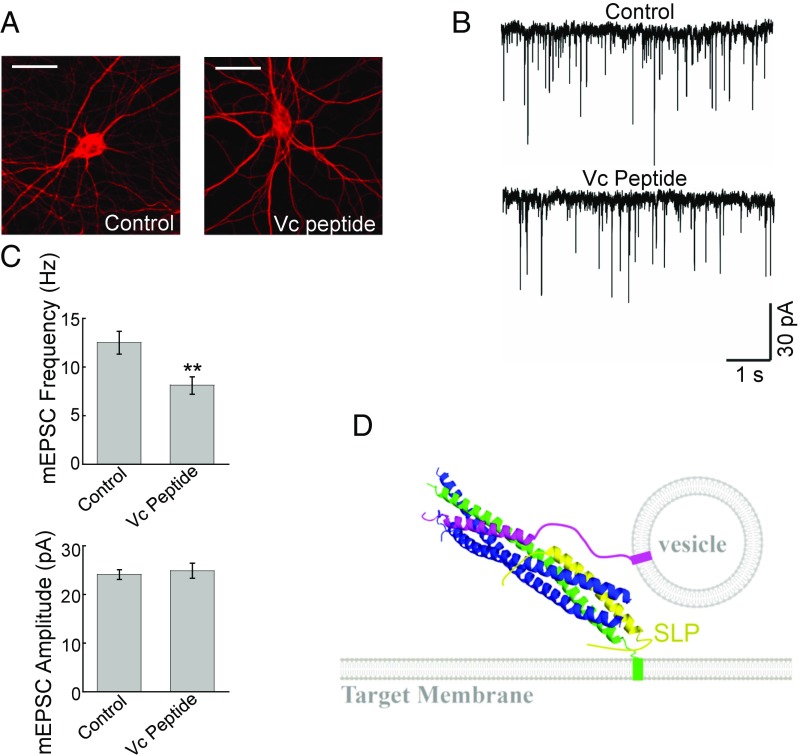
Fig. 7.
Expression of Vc peptide impairs the synaptic exocytosis in cultured neurons. (A) Representative immunofluorescence images showing cultured mouse cortical neurons expressing GFP (control) or GFP and Vc peptide. The cells were permeabilized and stained with anti-MAP2 antibodies. (Scale bars: 20 µm.) (B) Representative traces of miniature excitatory postsynaptic currents (mEPSCs) recorded in the control or Vc peptide-expressing cortical neurons. (C, Top) Summary graph of mEPSC frequency. (C, Bottom) Summary graph of mEPSC amplitudes. Numbers of neurons and independent cultures are listed in SI Appendix, Table S1. **P < 0.01. (D) Model illustrating the functional role of SLP in vesicle fusion reaction. SLP recognizes and restructures t-SNARE CTDs in the context of the half-zippered trans-SNARE complex. Green, Qa SNARE; blue, Qb and Qc SNAREs; magenta, v-SNARE (R-SNARE); yellow, SLP of the SM protein. For clarity, the rest of the SM protein is not shown. The model is based on the crystal structure of the synaptic exocytic SNARE complex (PDB ID code 1SFC).

