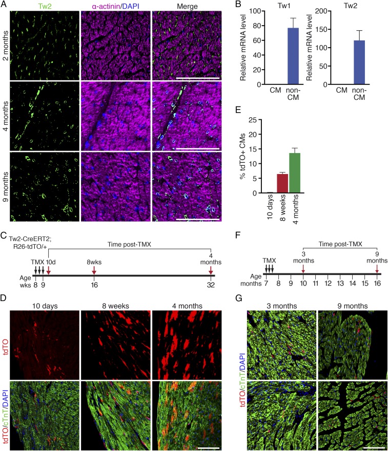
Fig. 1.
Progressive contribution of Tw2-tdTO+ cells to adult CMs. (A) Immunostaining of Tw2 (green) and α-actinin (violet) proteins on heart sections of 2-, 4-, and 9-mo-old wild-type mice. (Scale bars: 100 μm.) (B) Expression of Tw1 and Tw2 transcripts in CM-enriched and non–CM-enriched populations in adult mouse heart as detected by real-time RT-PCR (n = 3). (C) Schematic of TMX treatment. Tw2-CreERT2; R26-tdTO mice were injected with 1 mg of TMX on three alternative days at 8 wk of age. Mice were analyzed at 10 d, 8 wk, and 4 mo following the first TMX injection. (D) Progressive tdTO labeling of CMs at the indicated time points following TMX treatment. Sections were costained with cTnT (green) to detect CMs. (Scale bar: 100 μm.) (E) The percentage of Tw2-tdTO+ CMs in ventricles was quantified and averaged at the indicated time points. n = 3 mice for each time point. (F) Schematic of TMX treatment of aged Tw2-CreERT2; R26-tdTO mice. (G) Hearts from aged Tw2-CreERT2; R26-tdTO mice at the indicated times post-TMX treatment were costained with cTnT (green). (Scale bar: 100 μm.) Data in B and E are expressed as mean ± SEM.

