Fig. 3.
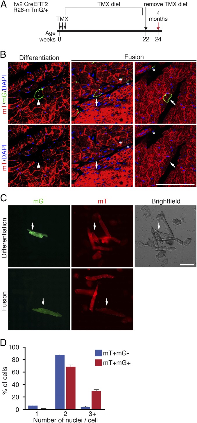
Tw2-tdTO+ cells fuse with CMs in adult hearts. (A) Schematic of TMX treatment of Tw2-CreERT2; R26-mT/mG mice. (B) Representative images of differentiation and fusion events detected in the hearts of Tw2-CreERT2; R26-mT/mG mice. (Left) The mG+ CMs no longer express the mT signal, indicating de novo CM formation (white arrowhead). (Center) The Tw2-mG+ CMs are mT− (white asterisks). (Right) The Tw2-mG+ CMs retain mT expression, indicating that Tw2-mG+ non-CMs fused with existing CMs (white arrow). Nuclei were stained with DAPI (blue). (Scale bar: 100 μm.) (C) CMs from ventricles of the Tw2-CreERT2; R26-mT/mG mice were dissociated and plated in culture. (Upper) A de novo CM that is mG+ but mT−. (Lower) Evidence of a fusion event in which the CM is mG+ and mT+ double positive. (Scale bar: 100 μm.) (D) Percent of cells that contain 1, 2, or >3 nuclei among all mT+ and mG− CMs or mT+ and mG+ CMs. n = 3 mice. Data in D are expressed as mean ± SEM.

