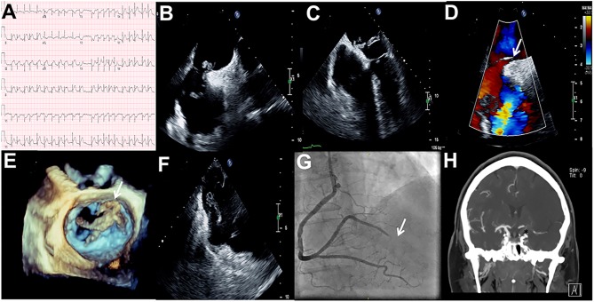
Figure 1.
(A) ECG showing an acute inferolateral ST-elevation MI; (B) TEE bicaval view revealing a highly mobile mass in the left and right atrium; (C) TEE four-chamber view showing a large thrombus across a PFO; (D) TEE bicaval with color Doppler showing a shunt across the interatrial septum; (E) 3D-TEE showing irregular shape of the thrombus; (F) TEE at lower-esophageal level showing a second smaller thrombus on the Eustachian valve; (G) CAG confirming 100% occluded distal right posterolateral artery; (H) CT angiography showing occluded internal carotid artery.

 This work is licensed under a