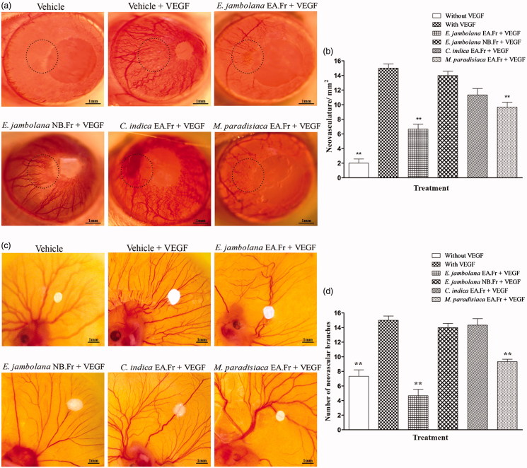
Figure 3.
Effects of plant extracts on neovasculature in the rat cornea and chick embryo CAM assays. (a) Representative photographs of VEGF-induced rat corneal neovascularization, plant extracts 20 μg/hydron polymer pellet were surgically implanted into the micro-pocket in the cornea of one eye. On day 7, the extent of neovascularization or inhibition was visualized and photographed under a dissection microscope. (b) Quantitative comparison of the number of neovascular vessels per mm2 was estimated. (c) Representative photographs of VEGF-induced neovascularization in shell-less CAM of chicken embryos. Plant extracts, 20 μg/filter disc was placed on the CAM of 6-day old chicken embryos. After 72 h of incubation, the area surrounding the filter disc was inspected for changes in neovascularization. (d) Quantitative comparison of the number of neovascular branches surrounding the plant extract containing filter paper discs. The data shown represent the results of experiments that were performed using a maximum of 5 eggs in each group. All quantitative data are presented as mean ± SEM of five independent counts; **p < 0.01.

