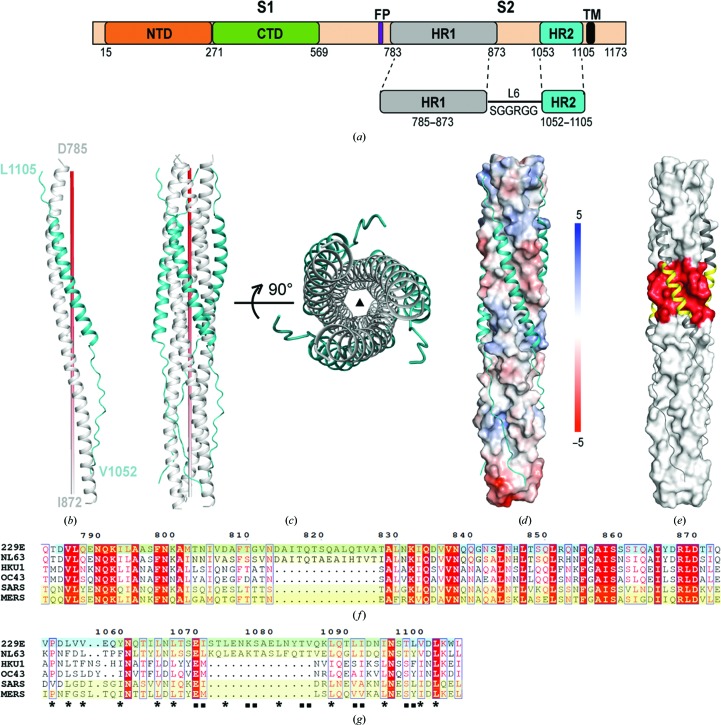
Figure 1.
Overall structure of the HCoV-229E fusion core. (a) Schematic diagram illustrating the design of the HR1-L6-HR2 fusion-core construct. (b, c) Monomeric (b) and trimeric (c) structures of the HR1-L6-HR2 fusion protein. The red pole (left panel) and filled triangle (right panel) in (c) represent the trimer threefold axis. (d) The packing of HR2 against the central hydrophobic core of HR1 as illustrated by a solvent-accessible surface rendering. The solvent-accessible surface is coloured according to the electrostatic potential, which ranges from +5 V (most positive, dark blue) to −5 V (most negative, dark red), with hydrophobic in white. (e) The HR2 helices are depicted as dark grey ribbons on the light grey surface of the HCoV-229E 3HR1 core to highlight the corresponding insertions in HR1 and HR2 in red and yellow, respectively. (f, g) Sequence alignment of the HR1 region (f) and HR2 region (g) of HCoV-229E, HCoV-NL63, HCoV-HKU1, HCoV-OC43, SARS and MERS. The HR1 and HR2 coverages of previous HCoV structures (PDB entry 2ieq for HCoV-NL63, PDB entry 1wyy for SARS and PDB entry 4njl for MERS) and the recently reported HCoV-229E structure (PDB entry 5zhy; Zhang et al., 2018 ▸) are depicted with a yellow background. The HCoV-229E structure presented here is denoted with a cyan highlight. The filled squares and stars underneath the residues highlight the conservation of residues in HR2 that are involved in hydrophobic interactions between HR1 and HR2 in the various HCoVs.

