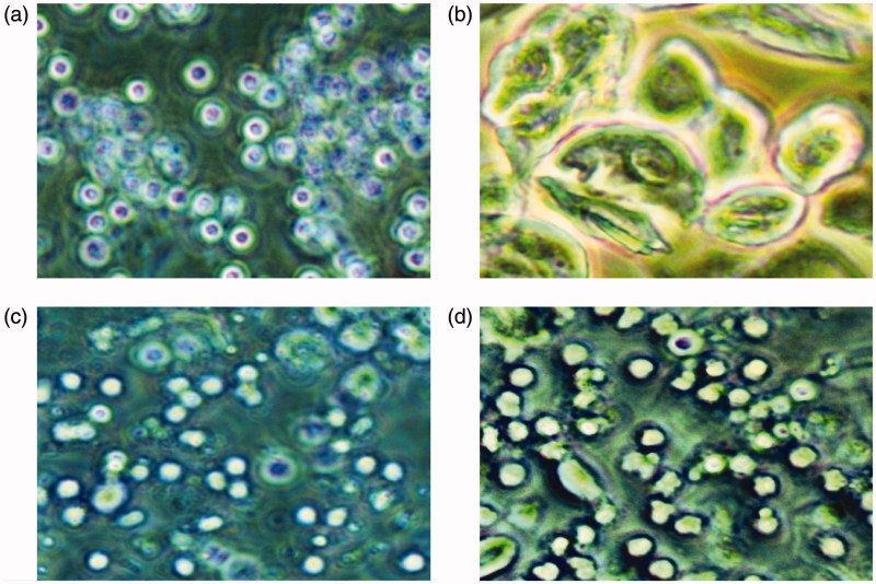
Figure 2.
The vaginal smears of rats with different stages of oestrous cycle in the control group. (a) The representative rat's vaginal smears from the control group in proestrous (200×). Oval nucleated epithelial cells, occasionally with a small number of keratinocytes, were detected. (b) The representative rat?s vaginal smears from the control group in estrous (200×). Epithelial keratinocytes with irregular shapes were detected; among which there was a small number of nuclear epithelial cells. (c) The representative rat?s vaginal smears from the control group in metestrous (200×). Irregular epithelial keratinocytes, nucleated epithelial cells, and leukocytes were detected. (d) The representative rat?s vaginal smears from the control group in diestrous (200×). A large number of leukocytes and a small number of nuclear epithelial cells were detected.

