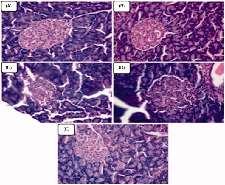
Figure 3.
(A–E) Represents the microphotographs of pancreatic tissues of normal control and experimental rats in each group (haematoxylin and eosin staining, 40×). (A and B) Normal control and normal control + geraniol (200 mg/kg body weight) treated rats showed normal appearing pancreatic exocrine glands and ducts with Islet of Langerhans. (C) Diabetic rats showing fatty infiltration and shrinkage of islet cells. (D and E) Pancreatic tissues from diabetic + geraniol (200 mg/kg b.w.) and diabetic + glyclazide (5 mg/kg b.w.) treated rats shows normal appearing pancreatic acini with absence of dilation and prominent hyperplastic of islets.

