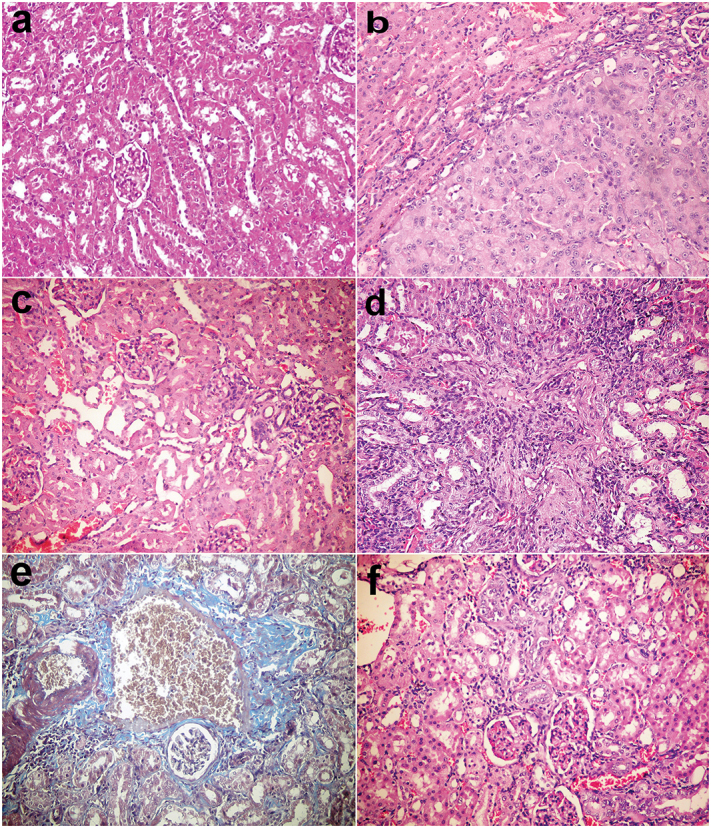
Figure 3.

Kidneys, rats (38th week). (a) Normal renal tissue in group I control group. (b) Tubular cell adenoma of solid pattern with compression of adjacent parenchyma in group V injected with DENA. (c) Minor histopathological alteration in group VI injected with DENA and treated with camel milk. (d) Mononuclear inflammatory cells infiltration in the interstitial tissue with thickening of glomerular and tubular basement membrane and fibroplasia in group VII injected with DENA and treated with cisplatin. (e) Bluish-stained periglomerular and interstitial fibroplasia (Massons’ trichrome stain). (f) Few mononuclear inflammatory cells infiltration with regenerated renal tubules in group VI injected with DENA and treated with camel milk and cisplatin. Haematoxylin and eosin stain 200×.
