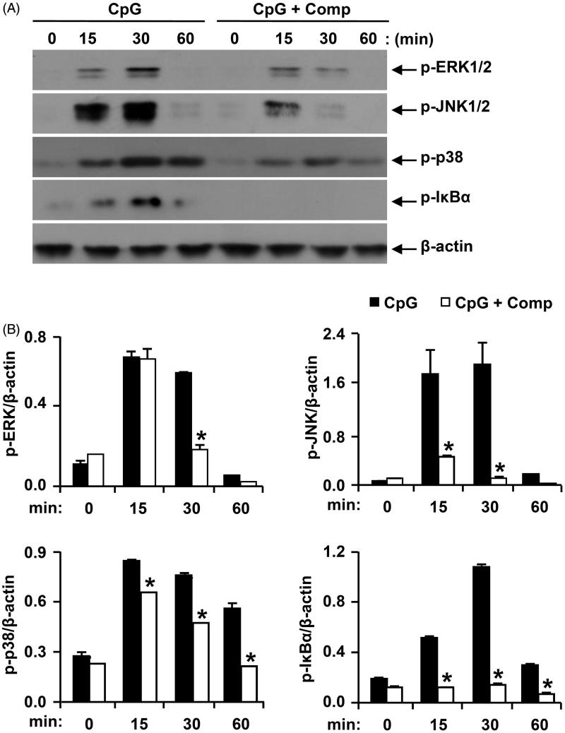
Figure 4.
Effects of comp on the phosphorylation of MAPK and IκBα by CpG DNA-stimulated BMDCs. (A) Cells were pretreated with or without comp (50 μM) for 1 h before stimulation with CpG DNA (1 μM). Total cell lysate was obtained at the indicated time intervals. Western blot analysis was performed on the cell lysate to assess phosphorylation of ERK, JNK, p38 and IκBα. β-Actin was taken as the loading control. Data are representative of three independent experiments. (B) Phosphorylation of ERK, JNK, p38 and IκBα protein expression was quantified using scanning densitometry, and the band intensities were normalized by that of β-actin. Comp: 3-hydroxy-4,7-megastigmadien-9-one. *p < 0.05 vs. comp-untreated cells in the presence of CpG DNA.

