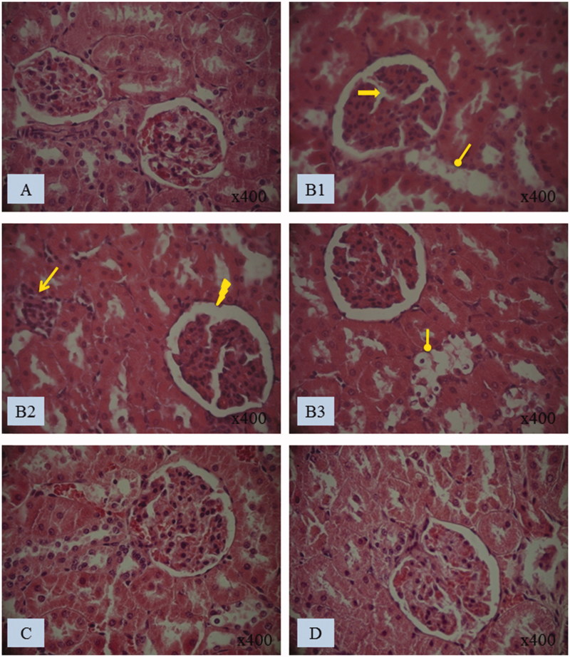
Figure 3.
Histological kidney sections of (A) control and (B1, B2 and B3) treated rats with penconazole, (C) N. retusa aqueous extract along with penconazole and (D) N. retusa aqueous extract. Optic microscopy: H&E (400×). Arrows indicate: Glomeruli fragmentation, necrosis of the epithelial cells lining the tubules, Bowman’s space enlargement, inflammatory leucocytes infiltration

