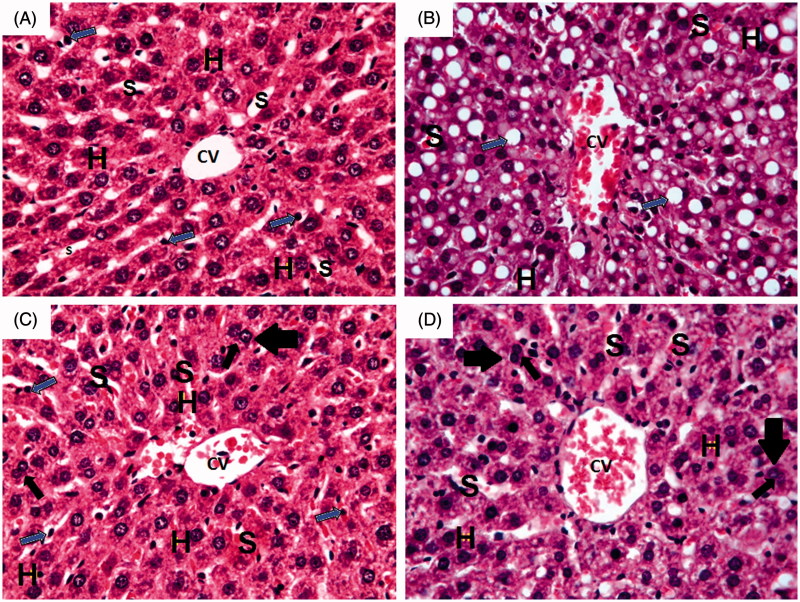
Figure 5.
Photomicrographs of liver section obtained from different groups (H&E; 400×), where (A) normal control group showing normal hepatic architecture with central vein (CV) and radiating cords of normal hepatocytes (H) with central rounded vesicular nuclei and prominent nucleoli. Hepatic cords are separated by blood sinusoids (S) lined with endothelium and von Kupffer cells (blue arrow); (B) ferrous sulphate group showing dilated congested central vein (CV) with congested blood sinusoids (S). Massive fatty infiltration of hepatocytes (H) with some hepatocytes acquired the signet ring appearance (blue arrow); (C) N-acetylcysteine plus ferrous sulphate group showing congested central vein (CV). Normal hepatocytes (H) are separated by slightly dilated congested blood sinusoids (S) with activated von Kupffer cells (blue arrow). Binucleated cells (black arrow) can be seen; (D) saponin plus ferrous sulphate group showing congested central vein (CV). Normal hepatocytes (H) are separated by slightly dilated congested blood sinusoids (S) with activated von Kupffer cells (blue arrow). Binucleated cells (black arrow) can be seen.

