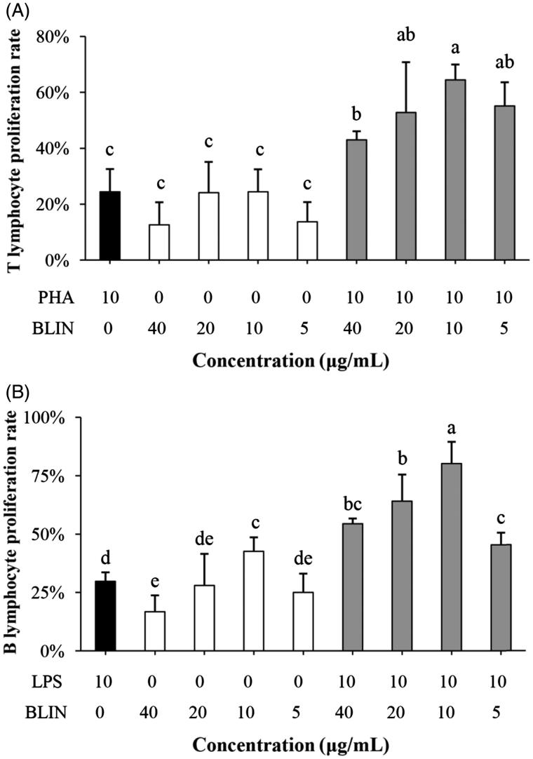
Figure 3.
Influence of BLIN on lymphocyte proliferation. PHA, as a control was used to stimulate the T lymphocyte. (A) BLIN at different working concentrations (20, 10, 5 and 2.5 μg/mL) stimulated T lymphocytes singly or co-stimulated with PHA, five repetitions per treatment. LPS, as a control, was used to stimulate the B lymphocyte. (B) BLIN at different working concentrations (20, 10, 5 and 2.5 μg/mL) stimulated B lymphocytes singly or co-stimulated with LPS, five repetitions per treatment. Statistical analyses were performed using Duncan’s multiple range tests. a–eBars in the figure without the same superscripts differ significantly (p < 0.05).

