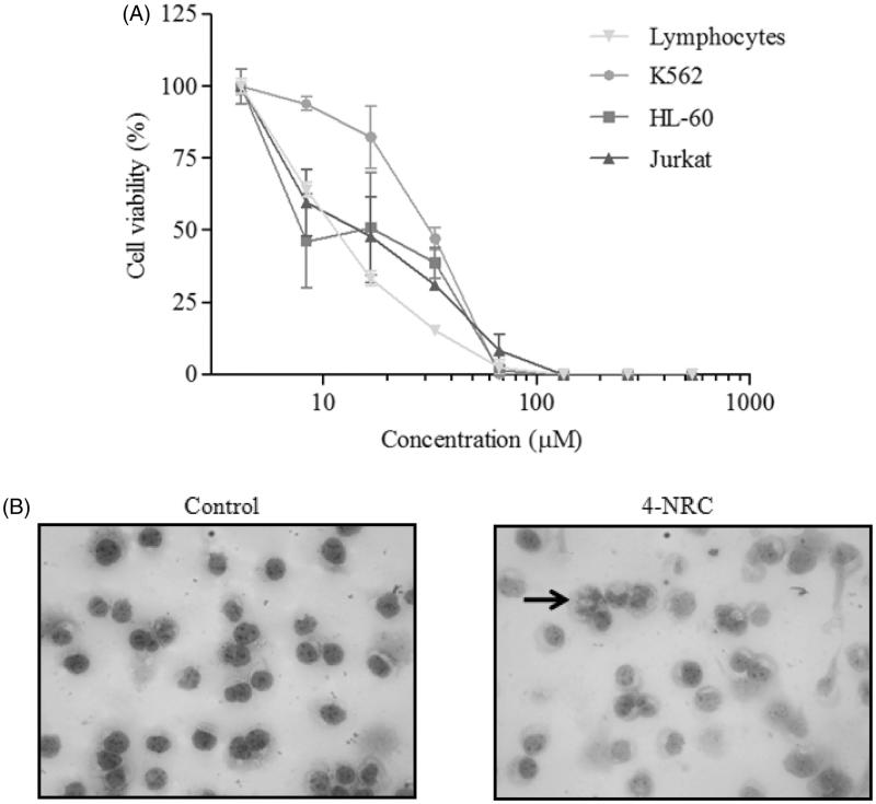
Figure 2.
Cytotoxic effects of 4-NRC in lymphocytes and leukemic cells. (A) Lymphocytes and K562, HL-60 and Jurkat cells (1 × 10⁶ cells/mL) were treated with different concentrations of 4-NRC (4.17–534.5 μM) for 24 h and cell viability was determined by MTT assay. Results represent the mean ± SD of three independent experiments in six replicates. (B) Morphological changes promoted by 4-NRC in K562 cells: control cells (2 × 10⁴/mL) stained with Giemsa dye showed normal nucleus while cells treated with 4-NRC (27 μM) for 24 h showed apoptotic nucleus (indicated by arrow). Representative photomicrographs (400 × magnification) of two independent experiments.

