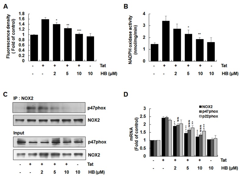
Fig. 3.
Effects of hindsiipropane B on HIV-1 Tat-mediated ROS generation and NADPH oxidase activation in CRT-MG cells. (A) Cells were pretreated with hindsiipropane B for 1 h and then stimulated with 50 nM HIV-1 Tat for 1 h. Cells were stained with DHE for 30 min. The levels of intracellular ROS probed with DHE were evaluated using an ELISA plate reader. (B) Cells pretreated with hindsiipropane B for 1 h were exposed to 50 nM HIV-1 Tat for 1 h. Cells were collected and mixed with 250 μM NADPH. NADPH depletion was observed by measuring reduction in absorbance at λ = 340 for 10 min. (C) Cells pretreated with hindsiipropane B for 1 h were exposed to 50 nM HIV-1 Tat for 1 h. Cell lysates were collected and immune-precipitated with gp91phox/NOX2 antibody, followed by Western blotting using a p47phox antibody to determine the levels of p47phox associated with gp91phox/NOX2. (D) Cells pretreated with hindsiipropane B for 1 h were stimulated with 50 nM HIV-1 Tat for 3 h. Total RNA was collected and assayed for mRNA levels of gp91phox/NOX2, p47phox, p22phox, and β-actin by quantitative RT-PCR. Data are shown as mean ± SD of three independent experiments. *P < 0.05, **P < 0.01, ***P < 0.001, ##P < 0.01, ###P < 0.001, ^^P < 0.01, and ^^^P < 0.001, as compared to the cells treated with HIV-1 Tat alone.

