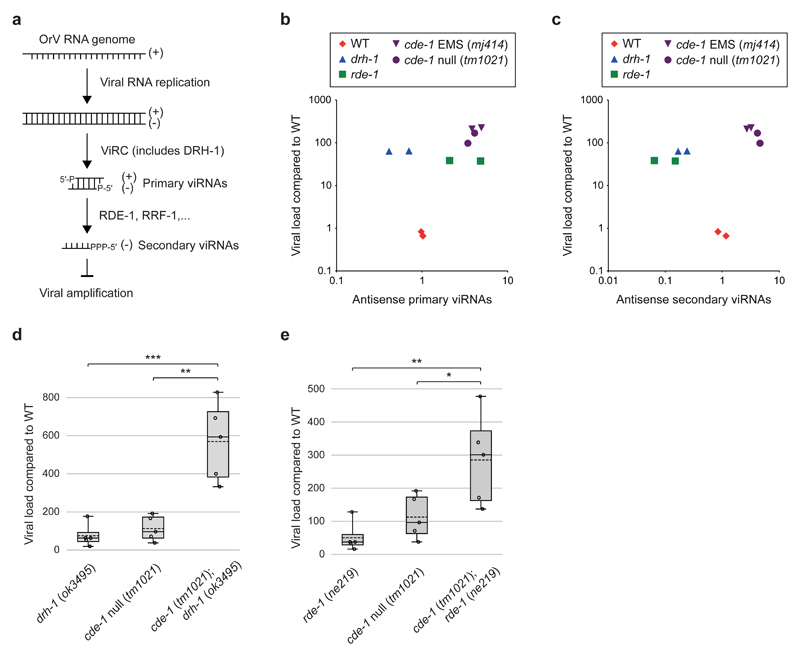
Figure 3. CDE-1 acts in parallel to antiviral RNAi.
a, Schematic of antiviral RNAi in C. elegans. Viral Recognition Complex (ViRC) includes DCR-1; DRH-1; RDE-4.
b, Comparison between the viral load and primary viRNA populations. Primary viRNAs (23-nucleotide long, from 5′ monophosphate RNA sequencing). Only antisense RNAs were considered to exclude potential viral genome degradation products. Dots: independent infection.
c, Comparison between the viral load and secondary viRNA populations. Secondary viRNAs (22-nucleotide long, starting with a G, from 5′ tri/monophosphate RNA sequencing). Samples as in b.
d and e, Viral load as measured by qRT-PCR of OrV RNA1 genome in adults two days after infection. Boxplots: whiskers from minimum to maximum; dots: independent infection; n=5. One-tailed student’s t-test: *** p<0.001, **p<0.01, *p<0.05. Samples as in b.

