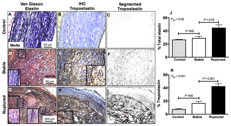
Figure 7. Histological analyses show accumulation of tropoelastin in rabbit rupture-prone compared with stable plaque.
A, D, G, Van Gieson elastin staining (dark purple indicates elastin fibers) shows the presence of organized elastin fibers in the media of control tissue and a net accumulation of elastin fibers in the intima of stable and rupture-prone plaque. B-C, E-F, H-I, Corresponding immunohistochemistry for tropoelastin (brown staining) shows lack of tropoelastin positive fibers in control aortas, upregulation in stable and deposition of a dense network of tropoelastin fibers in rupture-prone plaque. J-K, Quantification of total elastin and tropoelastin shows significantly higher tropoelastin in ruptured compared with stable plaque.

