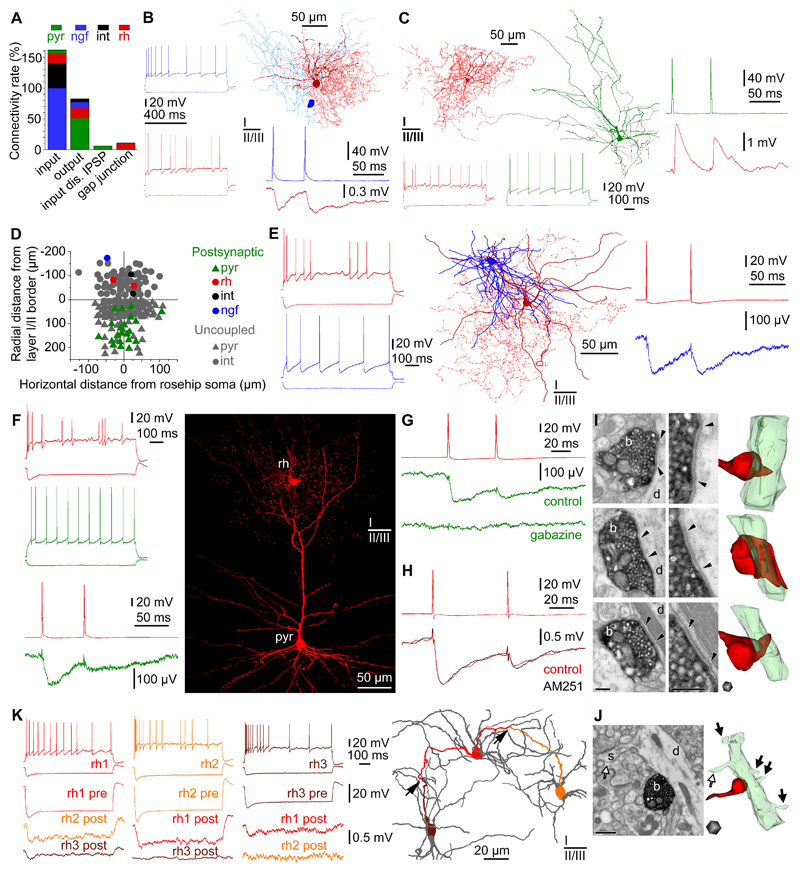
Figure 5. Connections of rosehip cells in the local microcircuit.
A, Distribution of local connections mapped in layers 1-3 between RCs (rh, red), pyramidal cells (pyr, green), NGFCs (ngf, blue) and other types of layer 1 interneuron (int, black) based on unbiased targeting of postsynaptic cells. RCs predominantly innervate pyramidal cells, receive monosynaptic EPSPs from layer 2-3 pyramidal cells, monosynaptic IPSPs from neurogliaform and other types of interneurons, however, IPSPs arriving from RCs were not encountered. In addition, RCs are interconnected by homologous electrical synapses (gap junctions). B, Example of a NGFC to RC connection. Left, Firing patterns of the presynaptic NGFC (blue) and the postsynaptic RC (red). Right, Anatomical reconstruction of the recorded NGFC (soma, dark blue; axon, light blue) and RC (soma and dendrites, burgundy; axon: red). Action potentials in the NGFC (blue) elicited slow IPSPs in the RC (red). C, Example of a pyramidal cell to RC connection. Left, Anatomical reconstruction and firing pattern of the presynaptic pyramidal cell (firing, soma and dendrites, green; axon, black) and the postsynaptic RC (firing, soma and dendrites, burgundy; axon, red). Right, action potentials in the pyramidal cell (green) elicited EPSPs in the RC (burgundy). D, Spatial distribution of coupled and uncoupled neurons tested as postsynaptic targets of RCs. Note the relative dominance of layer 2-3 pyramidal cells among neurons receiving input from RCs. E, The only RC to NGFC connection successfully tested for synaptic coupling. Left, Firing patterns of the presynaptic RC (burgundy) and the postsynaptic NGFC (blue). Middle, Anatomical reconstruction of the RC (soma and dendrites, burgundy; axon, red) and the NGFC (soma and dendrites, blue; axon not shown). Right, Action potentials in the RC (red) elicited slow IPSPs in the NGFC (blue). F, Example of RC to layer 3 pyramidal cell connections (n=16). Left, Firing patterns of the presynaptic RC (red) and the postsynaptic pyramidal cell (green). Action potentials in the RC (burgundy) elicited IPSPs in the pyramidal cell (green). Right, Confocal fluorescence image showing the recorded RC (rh) forming its axonal cloud in the tuft of the apical dendrite of the layer 2-3 pyramidal cell (pyr). G, Pharmacological characterization of a rosehip-to-pyramidal cell connection. Presynaptic spikes in the RC (red) elicited IPSPs in the layer 2-3 pyramidal cell (green) which could be blocked by application of gabazine (n=4, 10 µM). H, Functional test of presynaptic CNR1expression in RCs show the absence of modulation by the CNR1antagonist AM251 (n=4). Presynaptic spikes in the RC 1 (red, top) elicited IPSPs in the RC 2 (red, bottom). Application of AM251 (5 µM) had no effect on IPSPs (black). I, Representative electron microscopic images (left) and three-dimensional reconstructions (right, n=31) showing axon terminals (b, red) of biocytin filled RCs (n=3) targeting exclusively dendritic shafts (d, green) (100%, n=31). Synaptic clefts are indicated between arrowheads. Scale bars: 200 nm. J, Representative electron microscopic image (left) and three-dimensional reconstruction (right) of a biocytin filled RC bouton (b, red) targeting a pyramidal dendritic shaft (d, green) identified based on emerging dendritic spines (s, arrows). Scale bars: 500 nm. K, RCs form a network of electrical synapses. Top left, firing patterns of three RCs (red, rh1; orange, rh2; burgundy, rh3). Bottom left, Hyperpolarization of RC rh1 was reciprocally transmitted to RCs rh2 and rh3 confirming electrical coupling. Right, Route of the hyperpolarizing signals through putative dendro-dendritic gap junctions (arrows) between RCs rh1, rh2 and rh3 is shown by corresponding colors in the dendritic network of the three cells (gray).

