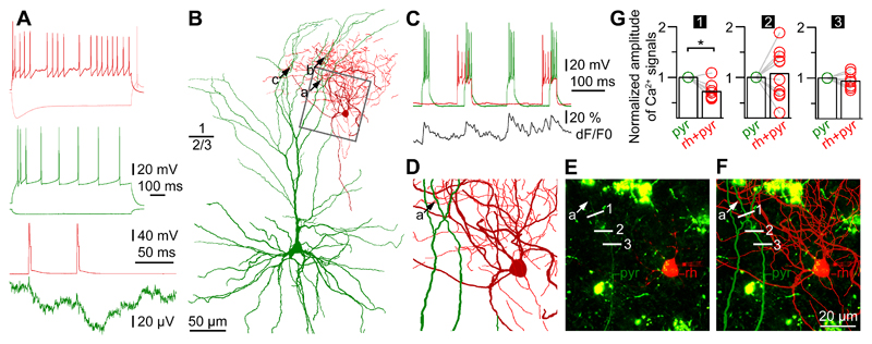
Figure 6. Human rosehip interneurons perform segment specific regulation of action potential backpropagation to apical dendritic tufts of pyramidal cells.
A, Top, Firing patterns of a presynaptic RC (burgundy) and a postsynaptic pyramidal cell (green). Bottom, Action potentials in the RC (burgundy) elicited IPSPs in the pyramidal cell (green). B, Anatomical reconstruction of the RC (soma and dendrites: burgundy; axon, red) and the layer 2-3 pyramidal cell (soma and dendrites, green; axon not shown). Presynaptic axonal boutons of the RC formed close appositions (a, b, and c) with three separate branches on the tuft of the pyramidal apical dendrite. C, Repetitive burst firing was triggered to initiate backpropagating Ca2+ signals in the pyramidal cell (green) while the output of the RC (red) was switched on and off timed prior and during every second pyramidal burst. Simultaneously, Ca2+ dynamics of the pyramidal apical dendritic tuft was measured at several locations and signals detected at location no.1 shown on panels E and F are shown in black. D, The area boxed in panel B shows the dendritic branch of the apical tuft of the pyramidal cell (green) with a putative synaptic contact (a) arriving from the RC. E, Confocal Z-stack image of the same area shown on panel D taken during paired whole cell recordings. The soma of the RC (rh, red), the dendrite of the pyramidal cell (pyr, green), the putative synaptic contact (a) arriving from the RC to the pyramidal cell and sites of line scans performed across the dendrite (1, 2 and 3) are indicated. Cytoplasmic lipofuscin autofluorescence characteristic to human tissue is seen as green patches. The experiment was repeated independently with similar results in n=4 cell pairs. F, Superimposition of the anatomical reconstruction of panel D and the confocal image of panel E. G, Normalized amplitudes of Ca2+ signals during pyramidal cell firing with and without coactivation of the RC detected at the three sites of line scans (1, 2 and 3) on the pyramidal dendrite. Rosehip input simultaneous with the backpropagating pyramidal action potentials was significant (p=0.02) in suppressing Ca2+ signals only at site 1 which was closest (8 µm) to the putative synapse between the two cells, no effect (n=10 trials, p=1 and p=0.27, respectively, two-sided Wilcoxon-test) of the RC was detected at sites 2 and 3 located at distances of 21 and 28 µm, respectively from the putative synaptic contact.

