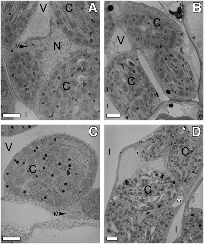
Fig 3. Transmission electron microscopy of mesophyll cells in uninjured and injured (21 hours post wounding) wild-type and ded1 leaves.
(A) Mesophyll cell in uninjured wild-type leaf tissue. (B) Mesophyll cell adjoining dead cells in injured wild-type leaf tissue. (C) Mesophyll cell in uninjured ded1 leaf tissue. (D) Mesophyll cell adjoining dead cell in injured ded1 leaf tissue. Note the swelling of the chloroplasts. Asterisks indicate location of cytoplasmic vacuoles. Bars = 1 μm. V = vacuole; C = chloroplast; I = intercellular space; M = mitochondrion; N = nucleus.

