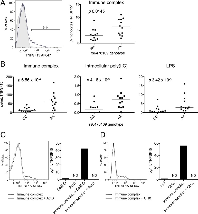
Fig 3. Genotype is associated with de novo TNFSF15 protein production in stimulated monocytes.
(A) An example of gating for TNFSF15+ monocytes after 4 hours immune complex stimulation is depicted (left). Black line = monoclonal anti-TNFSF15; grey shading = isotype control. Percentages of TNFSF15+ monocytes after immune complex stimulation are quantified for GG (n = 12) and AA (n = 12) homozygous individuals (right). (B) Soluble TNFSF15 was measured in supernatants of monocytes from GG (n = 12) and AA (n = 12) homozygous individuals after the indicated 4-hour stimulations by custom anti-TNFSF15 Bio-Plex assay. p values are from Mann-Whitney test. (C) Monocytes were stimulated with immune complex for four hours in the presence of actinomycin D (ActD) or dimethyl sulfoxide (DMSO) control. ND indicates none detected. (D) As (C) in the presence or absence of cycloheximide (CHX). (C) and (D) are representative of two independent experiments each.

