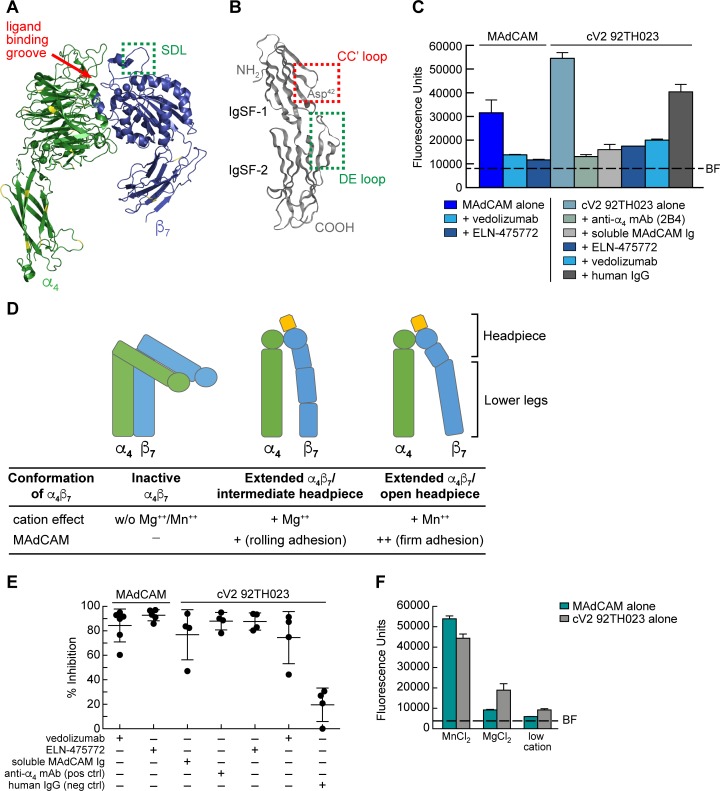
Fig 2. Inhibition of α4β7 -mediated cell adhesion to MAdCAM and a cyclic V2 peptide.
A) Ribbon diagram of a human α4β7 heterodimer headpiece (α4: green, β7: blue) (PDB ID: 3V4P). Yellow indicates sequence divergence in rhesus macaque. Ligand binding groove is highlighted by a red arrow, and the SDL is highlighted by a green box. B) Ribbon diagram of the two N-terminal IgSF domains of human MAdCAM (PDB ID: 1GSM). The MAdCAM CC′ loop of IgSF domain 1 is highlighted in the red box and the DE loop of IgSF domain 2 is highlighted in a green box. C) A representative result of RPMI8866 cells adhering to immobilized MAdCAM or a cV2 92TH023 peptide in the absence or presence of vedolizumab, an LDV mimetic ELN-475772, the anti-α4 specific mAb 2B4, soluble MAdCAM-Ig, or control human IgG, as indicated. Adhesion was determined at OD590nm and listed as fluorescence units (y-axis). Background fluorescence (BF) of RPMI8866 cell adhesion to blank wells is denoted by a dashed line. Adhesion to immobilized MAdCAM serves as a specificity control for α4β7 adhesion and human IgG is employed as a reagent control. Conditions are run in triplicate and error bars indicate standard error of the mean (SEM). D) Schematic of three conformations of the cell surface expression of the α4β7 heterodimer. The influence of divalent cations (Mg++ and Mn++) on the type of adhesion is listed below. E) Adhesion of RPMI8866 cells to either immobilized MAdCAM or cV2 92TH023 as in panel C. Average results from four or more independent experiments are shown. Y-axis indicates % inhibition relative to either MAdCAM or cV2 92TH023 in the absence of any inhibitor. Inclusion of inhibitors is indicated by a + sign below. Error bars indicate standard deviation (SD). F) Adhesion of RPMI8866 cells to immobilized MAdCAM or a cV2 92TH023 peptide in the buffers containing a low concentration of divalent cations, or high concentrations of MnCl2 or MgCl2. Adhesion was determined at OD590nm and listed as fluorescence units (y-axis). Conditions are run in triplicate and error bars indicate standard error of the mean (SEM). Background fluorescence (BF) of RPMI8866 cells to blank wells is denoted by a dashed line. Two additional replicate experiments are shown in S3 Fig.

