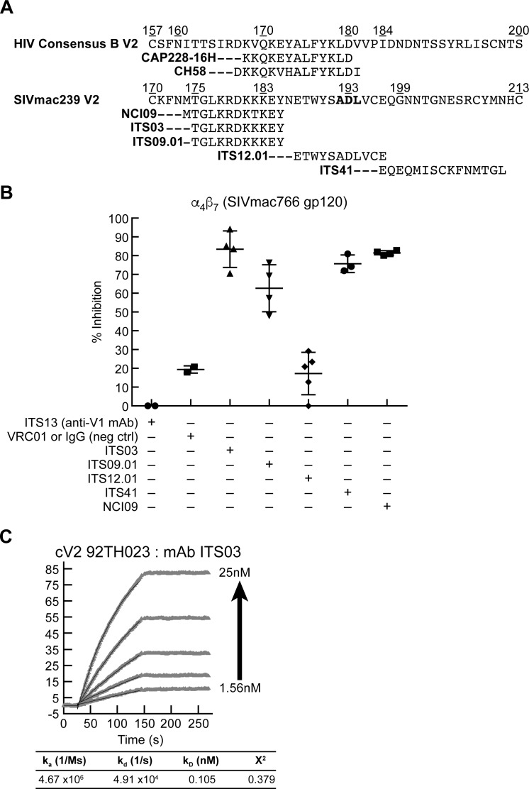
Fig 8. Inhibition of α4β7 adhesion by SIV V2 specific mAbs.
A) Alignment of the amino acid sequences of the V2 domains of HIV consensus B and SIVmac239, and the regions of the V2 containing epitopes recognized by V2 specific mAbs. B) Adhesion of RPMI8866 cells to DG SIVmac766 gp120 in the presence of SIV V2-specific mAbs: ITS03, ITS09.01, ITS12.01, ITS41, and NCI09. ITS13, a V1 mAb and VRC01, an HIV gp120 mAb are included as nonspecific mAb controls. Presence of each inhibitor indicated by a + sign below. Y-axis indicates % inhibition relative to adhesion in the absence of any inhibitor in three or more independent experiments. Error bars indicate SD. C) SPR analysis showing increasing concentrations of ITS03 mAb (2-fold increases from 1.56 nM-25 nM) binding to an HIV cV2 92TH023 coated surface. The parameters of binding kinetics are listed.

