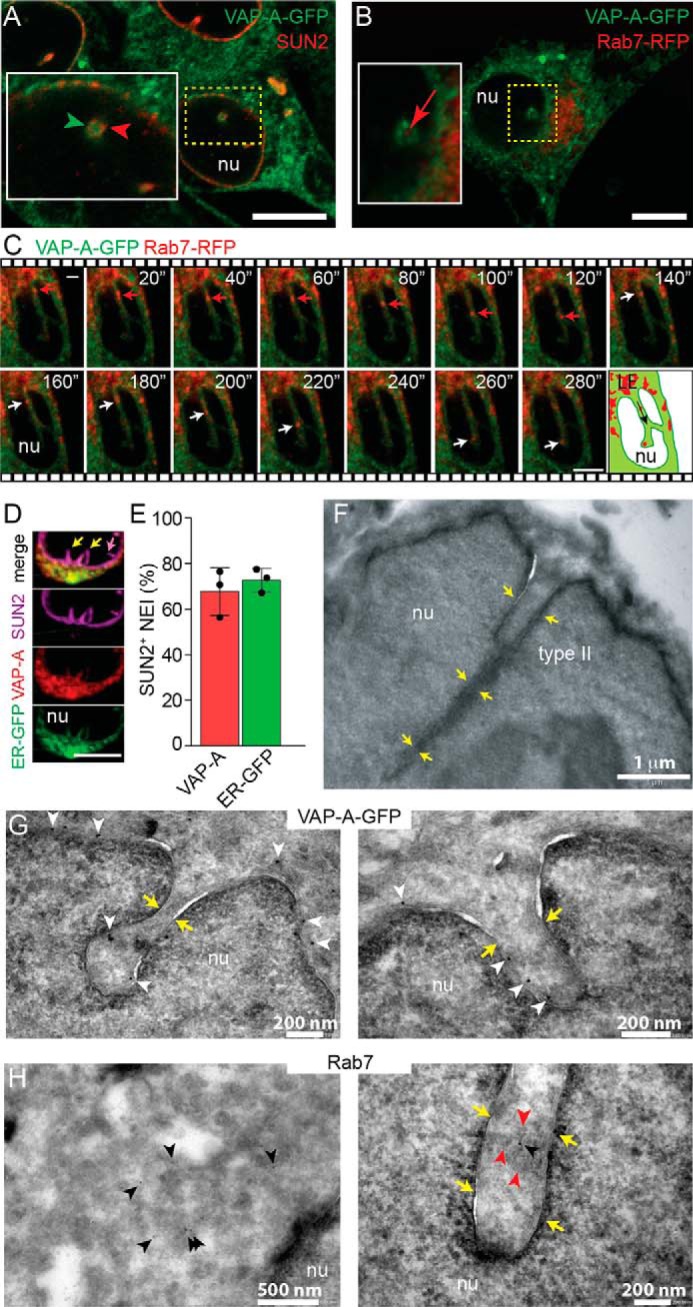
Figure 1.

VAP-A is associated with type II NEI containing late endosomes. A and B, VAP-A–GFP-transfected FEMX-I cells were immunolabeled for SUN2 (A) or infected with Rab7–RFP baculovirus (B) and analyzed by CLSM. Area of a cross-section of NEI is magnified. A single x-y optical section is presented. C, FEMX-I cells expressing VAP-A–GFP and Rab7–RFP were analyzed by time-lapse video microscopy. Elapsed time is indicated in the top right corner. Arrowheads show the localization of VAP-A–GFP in SUN2-immunolabeled NEI (A), and arrows indicate Rab7–RFP+ late endosomes in NEI (B and C). Cartoon illustrates the direction (arrow) of Rab7+ late endosomes (LE) in NEI. D, cells expressing ER–GFP marker were double-immunolabeled for VAP-A and SUN2. A three-dimensional (3D) reconstruction of two adjacent sections (0.4-μm each) is presented. Only part of the nucleus is shown. Note the absence (purple arrow) or presence (yellow arrows) of VAP-A and ER–GFP in SUN2+ type I and II NEI, respectively. Additional information is provided in Fig. S1. E, percentage of SUN2+ NEI (type II) containing VAP-A or ER–GFP is presented. The means ± S.D. are shown (n = 3). The average of each experiment, where more than 50 cells were evaluated, is indicated. F, EM of FEMX-I cells shows that type II NEI are not only superficial indentations of the nuclear envelope but also appeared as deep recesses (yellow arrows). G and H, immunogold labeling on ultrathin cryosections reveals the presence of VAP-A–GFP in deep NEI of FEMX-I cells expressing the fusion protein (G, white arrowheads), whereas Rab7 proteins (H, black arrowheads) are found in the cytoplasm (left panel) and NEI (right panel) of FEMX-I cells. The presence of membrane-bound organelles in NEI is indicated with red arrowheads. nu, nucleoplasm. Scale bars, 5 μm (A–D) or as indicated (F–H).
