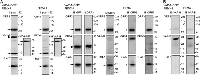
Figure 4.
Tripartite complex of VAP-A, ORP3, and Rab7 in NEI. A and B, detergent lysates prepared from transfected FEMX-I cells expressing VAP-A–GFP (VAP-A–GFP+ FEMX-I) or untransfected cells (FEMX-I) were subjected to immunoisolation (IS) with either anti-GFP antibody-coupled magnetic beads or anti-ORP3 (A) or anti-VAP-B (B) antibody followed by protein G-coupled magnetic beads as indicated above each lane. An aliquot of the input (1:50) and the entire bound fractions were probed for ORP3, VAP-A/B, and Rab7 by immunoblotting. Molecular mass markers (kDa) are indicated. The antibody used is indicated in the top left corner of the blot. Arrows indicate the protein of interest, and the arrowhead indicates VAP-A–GFP protein. Representative blots are shown (n = 3–6). Note that the VAP-A–GFP expression level is similar to endogenous protein in VAP-A–GFP+ FEMX-I cells, and almost no VAP-B is co-immunoisolated with ORP3 in contrast to VAP-A.

