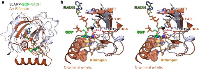
Figure 5.
Comparison between ScARP–GDP–NADH and Arr-rifampin (PDB code 2hw2). a, superimposed structures. Arr and rifampin are shown in brown and light brown, respectively. b, stereo view of the active sites. The nucleophilic nitrogen atom in GDP and oxygen atom in rifampin are shown as blue and red spheres, respectively. The electrophilic carbon atom in NADH is shown as a black sphere. Hydrophobic residues interacting with rifampin in the C-terminal α-helix are shown as stick and sphere models.

