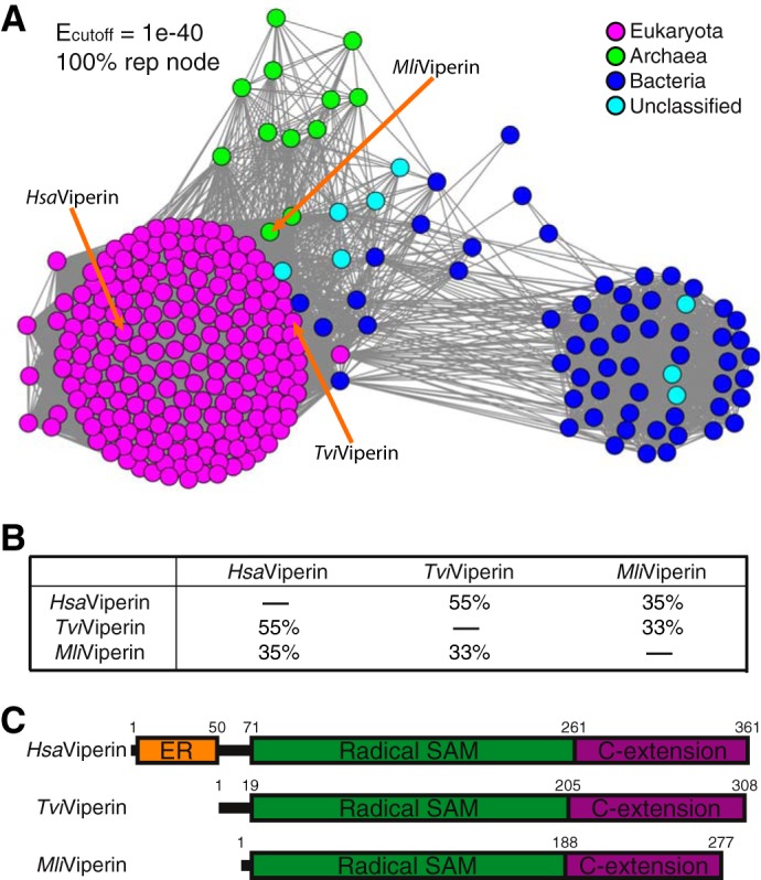
Figure 1.

Distribution and domain structure of viperin. A, SSN of viperin based on Blastp of HsaViperin. Three viperins employed in this study are marked with arrows. Tvi, Trichoderma virens; Mli, Methanofollis liminatans. B, pairwise amino acid sequence identities among the three viperins employed in this study. C, domain structures of these three viperins. The ER-associated domain (colored orange) present in HsaViperin is missing in both fungal TviViperin and archaeal MliViperin. C-extension, C-terminal extension.
