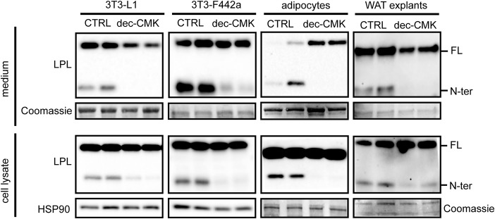
Figure 2.
LPL is cleaved by PCSKs in adipocytes. Shown are Western blots of cell culture medium (top panels) and cell lysates (bottom panels) of mature 3T3-L1 adipocytes, mature 3T3-F442a adipocytes, primary adipocytes differentiated from the stromal vascular fraction of adipose tissue from mice, and adipose tissue explants from mice that were treated with 50 μm dec-CMK for 9 h. Western blots were probed with antibodies against mLPL and HSP90 (as loading control). Coomassie Blue staining was performed as loading control for cell culture medium, primary adipocytes, and white adipose tissue (WAT) explants. FL, full-length; N-ter, N-terminal.

