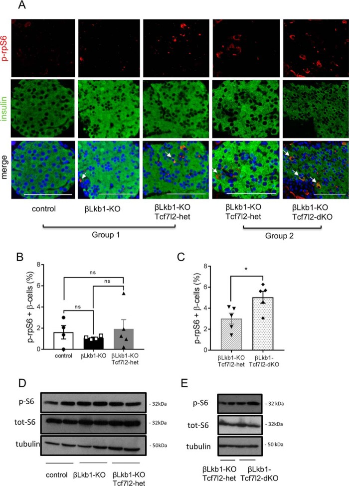
Figure 7.
Deletion of Tcf7l2 increases mTOR activity in Lkb1-null islets from males. A, representative immunofluorescence staining of pancreatic sections from random-fed 10-week-old males using rabbit anti-phospho-ribosomal protein S6 (Ser-235/236) (p-rpS6; 1:100; red) and guinea pig anti-insulin antibodies (1:200; red). White arrows represent phospho-rpS6 and insulin colocalization. Scale bars, 100 μm. B and C, quantification of phospho-rpS6–positive staining of total β cells per islet based on 15–20 islets per pancreas from n = 4–5 mice/genotype. White bars, control; black bars, βLkb1-KO; gray bars, βLkb1-KO-Tcf7l2-het (B), gray hatched bars, βLkb1-KO-Tcf7l2-het (C); and black dotted bars, βLkb1-Tcf7l2-dKO. D and E, mouse pancreatic islets were isolated from animals of group 1 (D) and group 2 (E). After an overnight incubation in 11 mmol/liter glucose in RPMI 1640 medium, isolated islets were collected, and lysates from 125 islets were analyzed by immunoblotting with anti-phosphorylated (p-S6) and total ribosomal protein S6 (tot-S6) (Ser-235/236) and anti-tubulin for group 1 (D) and group 2 (E). Error bars represent the mean ±S.E.; *, p < 0.05; ns, not significant.

