
Figure 1. Temporal and spatial distribution of NF-α1 in embryos.
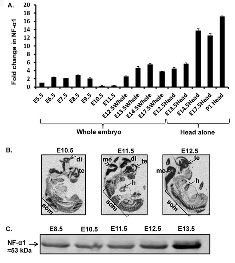
(A) Bar graphs show NF-α1 mRNA expression in E6.5 embryos to postnatal day1 (P1) (head only) relative to E5.5 embryos. Values are mean ± SEM; N=3, n=3 per embryo stage. (B) In situ hybridization indicates NF-α1 mRNA highly expressed in embryonic brain especially di (diencephalon), te (telencephalon), som (somites), me (mesencephalon), and h (heart) (N=3). (C) NF-α1 was immunoprecipitated with polyclonal rabbit anti-NF-α1 Ab from whole embryos (E8.5–11.5) or embryo head (E12.5& E13.5) and probed with mouse anti-NF-α1 Ab. NF-α1 protein is detectable at early embryonic stages (E8.5, 10.5, 11.5, 12.5 & 13.5).