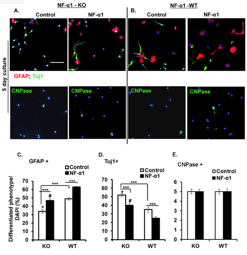
Figure 5. NF-α1-KO/NF-α1-WT generated neural stem/progenitors indicate a modulatory role of NF-α1 in altering the differentiation cell phenotype.

Neural stem/progenitors generated from NF-α1-KO and NF-α1-WT were grown for 5 days in differentiation media and treated with or without NF-α1 on day 0. At the end of day 5 the cells were stained and analyzed for GFAP, β-III tubulin and CNPase positive cells. Representative ICC pictures (x20) from NF-α1-KO (A) and NF-α1-WT (B) cells. Scale bar = 100 µm. Left and right side panels indicate controls and NF-α1 treated cells. C, D & E show bar graph analysis comparing between NF-α1-KO and NF-α1-WT for individual cell (GFAP+, Tuj1+ & CNPase+) phenotypes. Differentiated NPCs derived from NF-α1-KO (control) showed a significant ~30% decrease in GFAP+ cells (C) and ~34% increase in Tuj1+ cells (D) when compared to NF-α1-WT controls. Irrespective of NF-α1-KO and NF-α1-WT cultures, NF-α1 treatment increased GFAP+ cells and decreased Tuj1+ cells (C, D). +p=0.03, #p<0.001, ***p<0.0001, N=3, the values represent the mean ± SEM, one way ANOVA (Tukey’s multiple comparison test) and t test.
