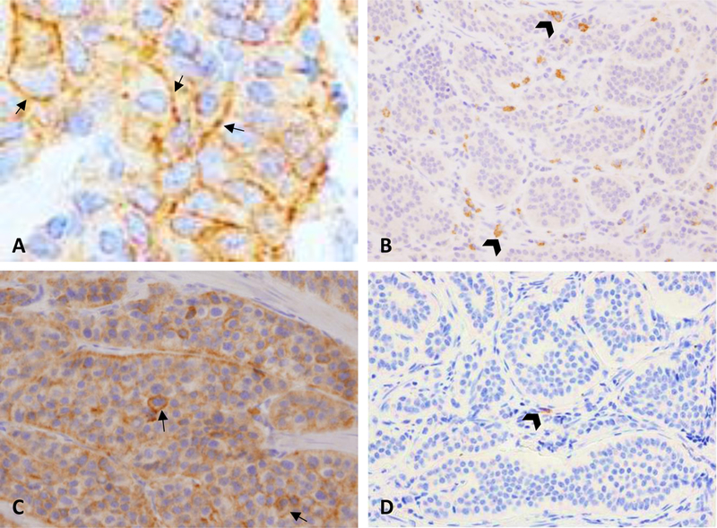
FIGURE 1. PD-L1, PD-L2 and PD-1 in tumor and stromal cells.

A, Membranous expression of PD-L1 in pNET. High expression was rare and found in only 7% of cases. B, High expression of PD-L1 in immune stromal cells (head arrows) was found in only a single case of SINET. C, High expression of PD-L2 in pNET tumor cells. Expression was predominantly cytoplasmic with approximately 5% of cells also showing membranous expression (arrows). D, Representative low density expression of PD-1 in immune stromal cells in SINET (head arrow).
