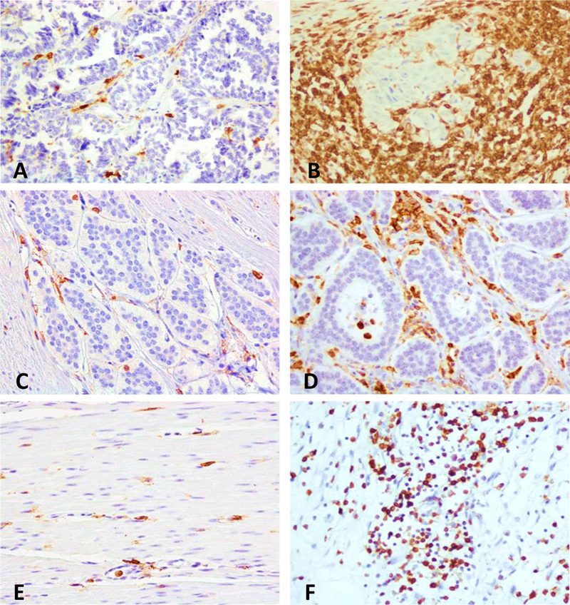
FIGURE 3. Representative intra and extratumoral lymphocytic infiltrates in pNET and SINET.

CD45RO+ T-cells are shown in the intratumoral (A-D) and extratumoral compartments (E, F). A, C, and E show a low density of CD45RO+ cells, in the intratumoral compartment of a pNET and SINET, and in the extratumoral compartment of a SINET respectively. B, D, and F show a high density of CD45RO+ cells, in intratumoral compartment of a pNET and SINET, and in the extratumoral area of a pNET, respectively.
