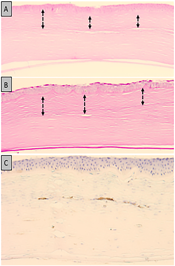
Figure 3.

Photomicrograph of human cornea 16 months after rose bengal PDAT for Fusarium keratitis. Anterior stromal scarring (double arrow) with disorganization of the collagen bundles present to a depth of 220 μm. (A: Hematoxylin and eosin; original magnification × 200) (B: Periodic acid–Schiff ; original magnification × 200) C: Negative alpha-smooth muscle actin staining within the stromal cells and focal positivity within blood vessels.
