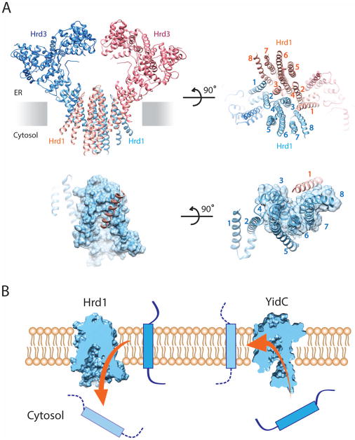
Figure 2. Structure of a Hrd1/Hrd3 complex.
(A) Model of Hrd1 bound to the luminal domain of Hrd3, based on cryo-EM single-particle analysis [38]. The upper left panel shows a cartoon of the Hrd1/Hrd3 dimer, with the Hrd1 molecules in light blue and salmon, and the Hrd3 molecules in dark blue and red. The upper right panel shows a view from the cytosol. The lower left panel shows a space-filling model of the funnel of one Hrd1 molecule together with TM1 of the other. The lower right panel shows a view from the cytosol. (B) The left panel shows a cut through a space-filling model of Hrd1. Hrd1/Hrd3 allows an ERAD-M substrate to move into the cytosol (arrow). The right panel shows a cut through a space-filling model of the bacterial YidC protein, which allows TM segments to move from the cytosol into the membrane (arrow).

