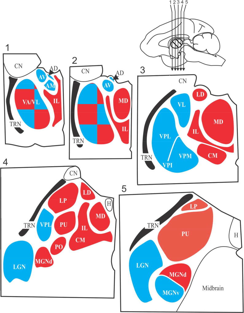
Figure 2.
Schematic view of five sections through the thalamus of a monkey. The sections are numbered 1 through 5 and were cut in the coronal planes indicated by the arrows in the upper right mid-sagittal view of the monkey brain. The major thalamic nuclei in one hemisphere for a generalized primate are shown. First order nuclei are shown in blue and higher order nuclei are shown in red; note that VA/VL is a nuclear complex that appears to be a mosaic of first and higher order regions. Abbreviations: AD anterodorsal nucleus; AM anteromedial nucleus, AV anteroventral nucleus; CM, centermedian nucleus; CN caudate nucleus (not a part of the thalamus); H, habenular nucleus; IL, intralaminar (and midline) nuclei; LD, lateral dorsal nucleus; LGN, lateral geniculate nucleus; LP, lateral posterior nucleus; MGN, medial geniculate nucleus; PO, posterior nucleus; PU pulvinar; TRN, thalamic reticular nucleus; VA, ventral anterior nucleus; VL ventral lateral nucleus; VPI, VPL, VPM, are the inferior, the lateral and the medial parts of the ventral posterior nucleus or nuclear group.

