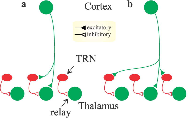
Figure 4.
Schematic circuit diagrams illustrating two possible scenarios for the organization of layer 6 corticogeniculate input to relay neurons and inhibitory interneurons in the thalamus. a. One scenario where corticogeniculate neurons provide monosynaptic excitation to retinotopically aligned relay cells and aligned inhibitory cells (TRN cells or local interneurons) that, in turn, inhibit the same relay cell receiving monosynaptic excitation. b. An alternative scenario where corticogeniculate neurons provide monosynaptic excitation to retinotopically aligned relay cells and non-aligned inhibitory neurons. In this scenario, relay cells receive monosynaptic excitation from retinotopically aligned corticogeniculate neurons (relay cell 2) and disynaptic inhibition from non-aligned corticogeniculate neurons (relay cells 1 and 3).

