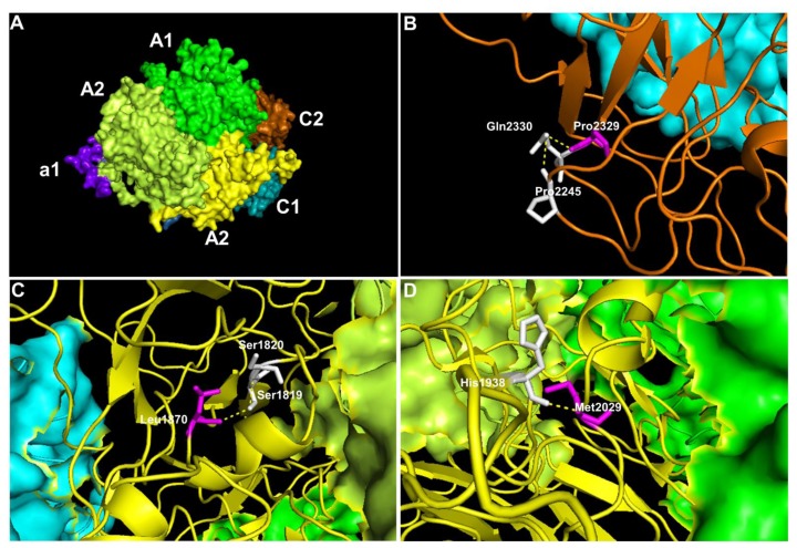
Figure 1.
The representative model of factor VIII protein showing the affected amino acids by missense mutations. The visualisation of affected amino acids by missense mutations based on factor VIII protein (PDB:2R7E). A) The localisation of the domains in the factor VIII, B) Position of Pro2329 in the C2 domain C) Leu1870 in the A3 domain and D) Met2029 in the A3 domain. The visualisation of the whole structure of factor VIII as a surface model with colour coding that represents A1 domain (green), a1 domain (purple), A2 domain (lime), A3 domain (yellow), C1 domain (cyan) and C2 domain (orange). Except when the domain is affected, the region is visualised as a ribbon model. In this ribbon model, the affected residue (magenta), the neighbouring amino acids (white), and the hydrogen bonds (yellow dotted lines) are highlighted.

