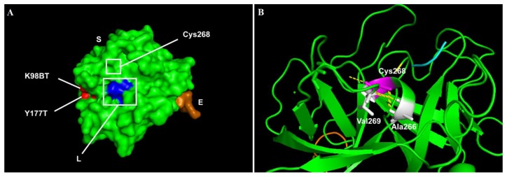
Figure 2.
The representative model of factor IX protein showing the affected amino acids by missense mutations. The visualisation of affected amino acids by missense mutations based on factor IX protein (PDB:2WPL) of double-mutant. A) Visualisation of the selected structure of factor IX protein as a surface model with colour-coding that represents S chain, containing Peptidase S1 domain (green), E chain containing EGF2 domain (orange) and L chain domain (blue). Original mutants’ residues from the crystal structure (2WPL) are in red. B) The affected domain of Peptidase S1 is visualised as a ribbon model, with the affected residue (Cys268, magenta), the neighbouring amino acids (white), and the hydrogen bonds (yellow dotted lines) are highlighted.

