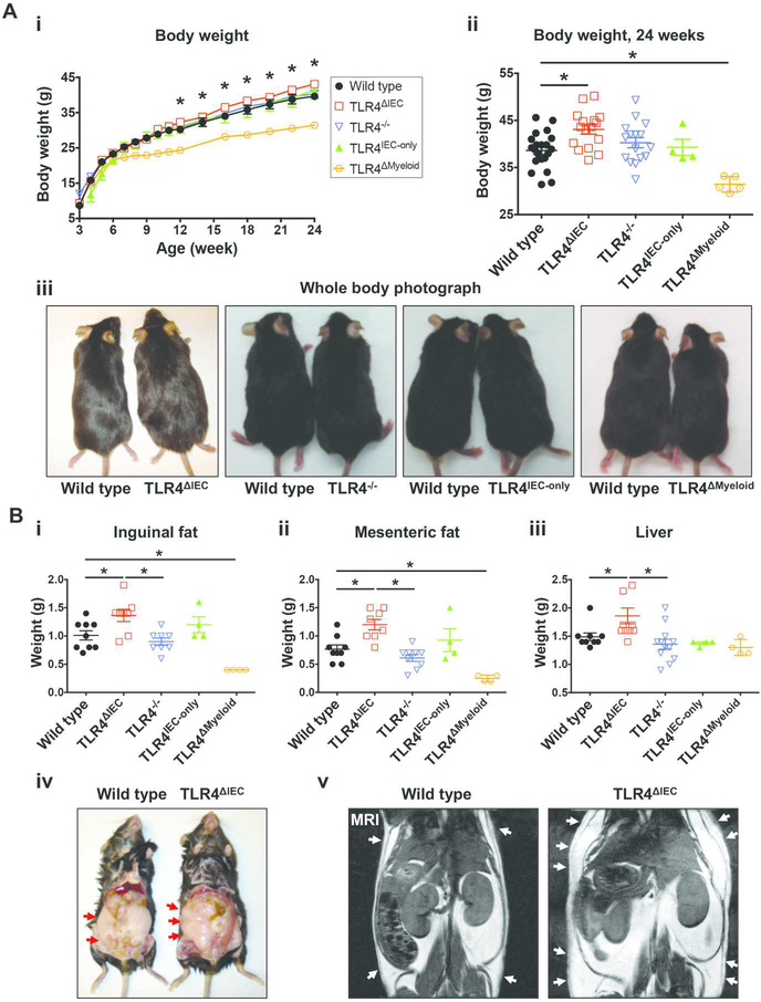
Figure 1. Deletion of the lipopolysaccharide receptor TLR4 from intestinal epithelial cells leads to obesity in mice.
A: i: Determination of body weight between 3 and 24 weeks of wild type (n=33), TLR4ΔIEC (n=31), TLR4−/− (n=31), TLR4IEC-only (n=6) and TLR4ΔMyeloid (n=8) mice who were fed standard chow; ii: body weight at 24 weeks for the wild type (n=21), TLR4ΔIEC (n=16), TLR4−/− (n=16), TLR4IEC-only (n=4) and TLR4ΔMyeloid (n=5) mice is shown. Data are represented as mean ± SEM; *p<0.05 wild type vs TLR4ΔIEC mice at the indicated time point; each symbol represents a separate mouse; iii: representative photographs of wild type, TLR4ΔIEC, TLR4−/−, TLR4IEC-only and TLR4ΔMyeloid at 24 weeks of age is shown revealing the significantly larger size of the TLR4ΔIEC mice compared with wild type mice. B: Weight of inguinal fat (i), mesenteric fat (ii) and liver (iii) of wild type (n=9), TLR4ΔIEC (n=8 to 10 as indicated), TLR4−/− (n=8 to 12 as indicated), TLR4IEC-only (n=4) and TLR4ΔMyeloid (n=4) mice fed standard chow at the age of 24 weeks. Data are represented as mean ± SEM; *p<0.05 for the indicated comparison; each symbol represents a separate mouse; iv-v: representative photograph (iv) and micro-MRI (v) revealing increased abdominal fat in the TLR4ΔIEC as compared with wild type mice at 24 weeks of age; arrows show the deposition of adipose tissue in the subcutaneous tissue.

