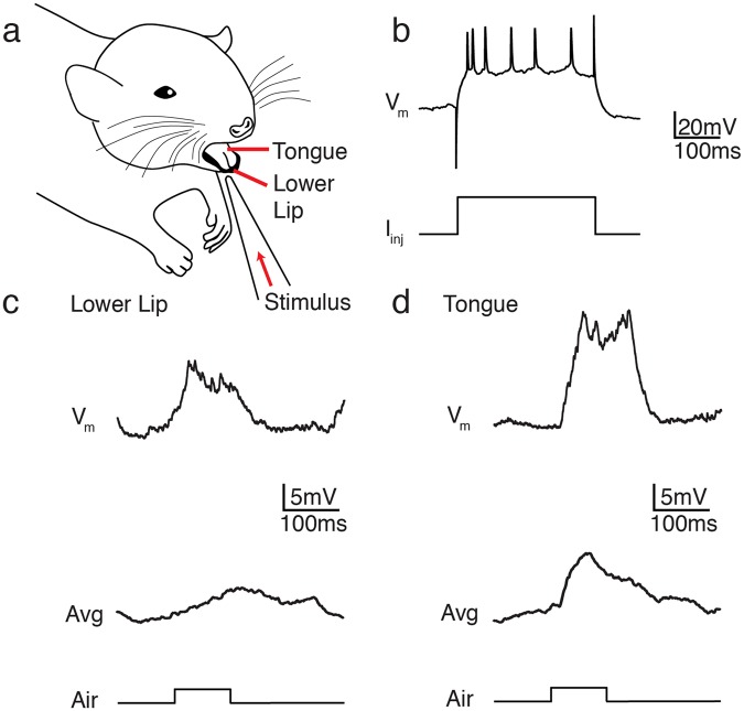
Figure 1.
Stimulation of intra-oral receptive fields in somatosensory cortex (S1). (a) Stimulated area of an anaesthetized rat. The lower lip or tongue was stimulated with an air puff generated by a computer-triggered airflow controller. (b) Representative voltage trace (Vm) from a whole-cell patch clamp recording from an S1 cortical neuron during current injection (Iinj). (c,d) Example whole-cell recordings from a representative S1 cortical neuron showing a single trial response (top) and averaged response (bottom; n = 19 trials) to air puff stimulation of the lower lip (c) or tongue (d). Timing of stimulus delivery is indicated below each trace.

