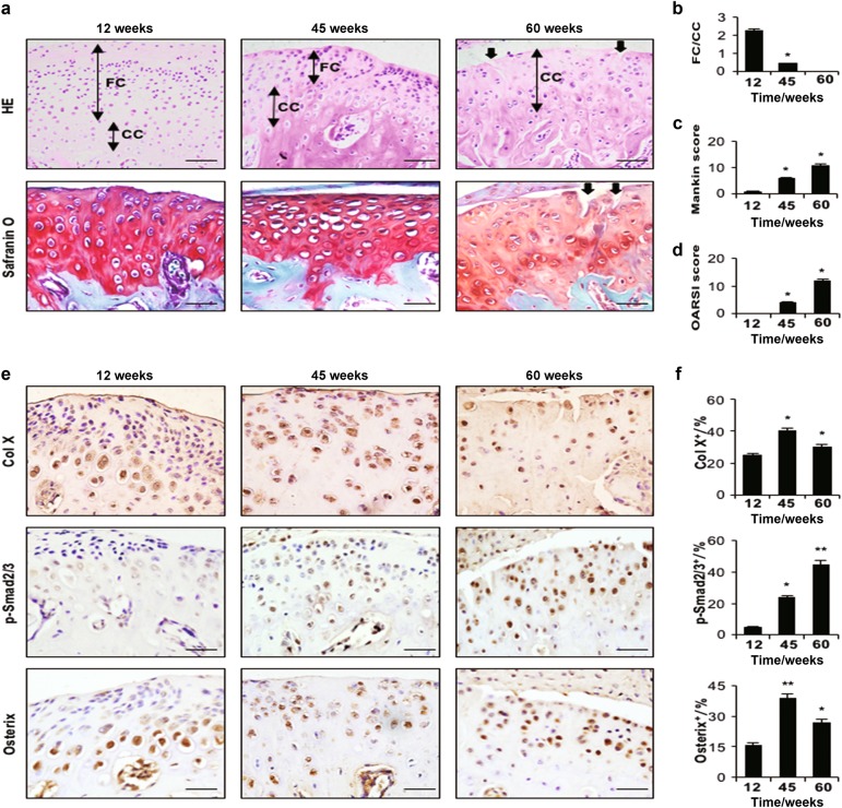
Fig. 2.
Condylar cartilage degeneration in ageing mice at different time points (12 wks, 45 wks and 60 wks). a HE (top) and Safranin O and fast green (bottom) staining analyses of glycosaminoglycan (red), black arrow indicates a crack. Mandibular condylar cartilage cell layers (FC fibrocartilage layer, CC calcified cartilage layer). b FC/CC, c Mankin and d OARSI scores of ageing mice. e Immunohistochemical analyses of Col X, p-Smad2/3 and Osterix (brown) in the mandibular condylar cartilage of ageing mice. f Col X-, p-Smad2/3- and Osterix-positive cells were counted in the cartilage layer. Scale bars = 20 µm. n = 6 per group. *P < 0.05, **P < 0.01 one-way ANOVA followed by Tukey’s test. All data are expressed as the mean ± s.d

