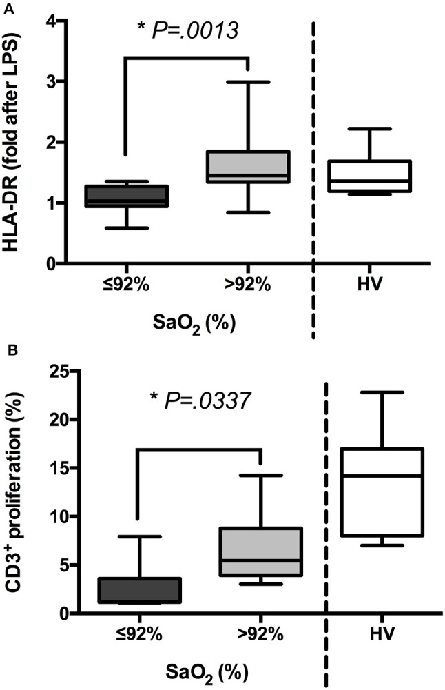
Figure 2.

The groups of patients with sepsis exhibit different states of activation after ex vivo challenge. (A) Blood samples from patients with sepsis (n = 85) and healthy volunteers (HV, n = 15) were stimulated or not with LPS (5 ng/mL, 3 h) ex vivo. Then, mean intensity of fluorescence (MIF) of HLA-DR on the gate of CD14+ cells was analyzed by FACS. Folds after LPS challenge are shown in patients classified according to their oxygen saturation and HV. (B) PBMCs were isolated from patients with sepsis (n = 85) and HV (n = 15), labeled with CFSE and stimulated or not with PWD (2.5 μg/mL) for 5 days. Then, proliferation of CD3+ cells was analyzed by FACS. Percentages of proliferation are shown in patients classified according to their oxygen saturation and HV *p < 0.05 using a Student's t-test.
