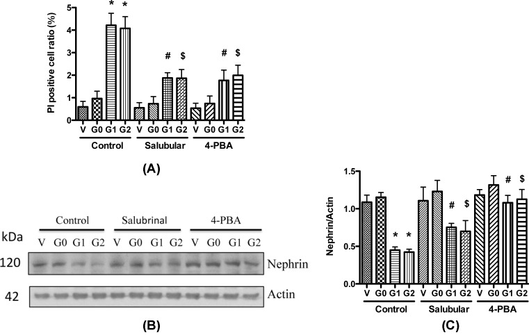
Figure 7. ER stress inhibitors attenuate APOL1 risk variants caused podocyte injury.
(A) Differentiated HPs (APOL1-Vec/HPs, G0/HPs, G1/HPs, and G2/HPs) were treated with Salubrinal (2.5 μM) or 4-PBA (1 mM) for 48 h, and cell lysates were collected for PI staining. Necrotic cells were counted, and the cell ratio was calculated. *, #, and $, P<0.05 in comparison with APOL1-G0, APOL1-G1, and APOL1-G2 in control group, respectively. (B,C) Differentiated HPs (APOL1-Vec/HPs, G0/HPs, G1/HPs, and G2/HPs) were treated with Salubrinal (2.5 μM) or 4-PBA (1 mM) for 48 h, and cell lysates were collected for Western blotting to detect nephrin protein. Representative gels are shown in (B). The protein bands were scanned and the acquired images were analyzed using the public domain NIH image program for data quantitation. Expression of nephrin (C) were normalized to β-actin. Data are presented as fold of control expression. *, #, and $, P<0.05 in comparison with APOL1-G0, APOL1-G1, and APOL1-G2 in control group, respectively.

